


4 - 7 September 2026
Prague | Czech Republic
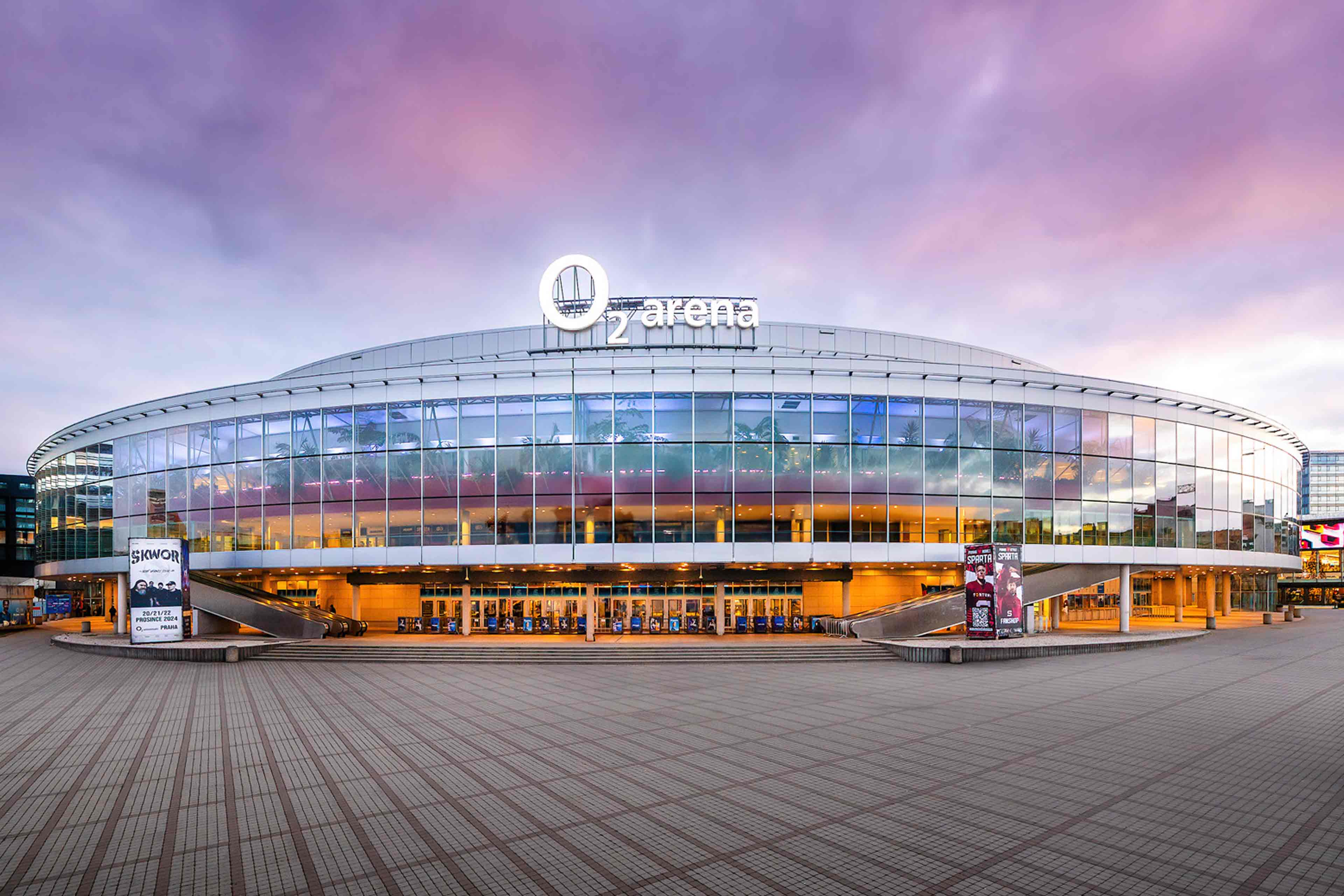
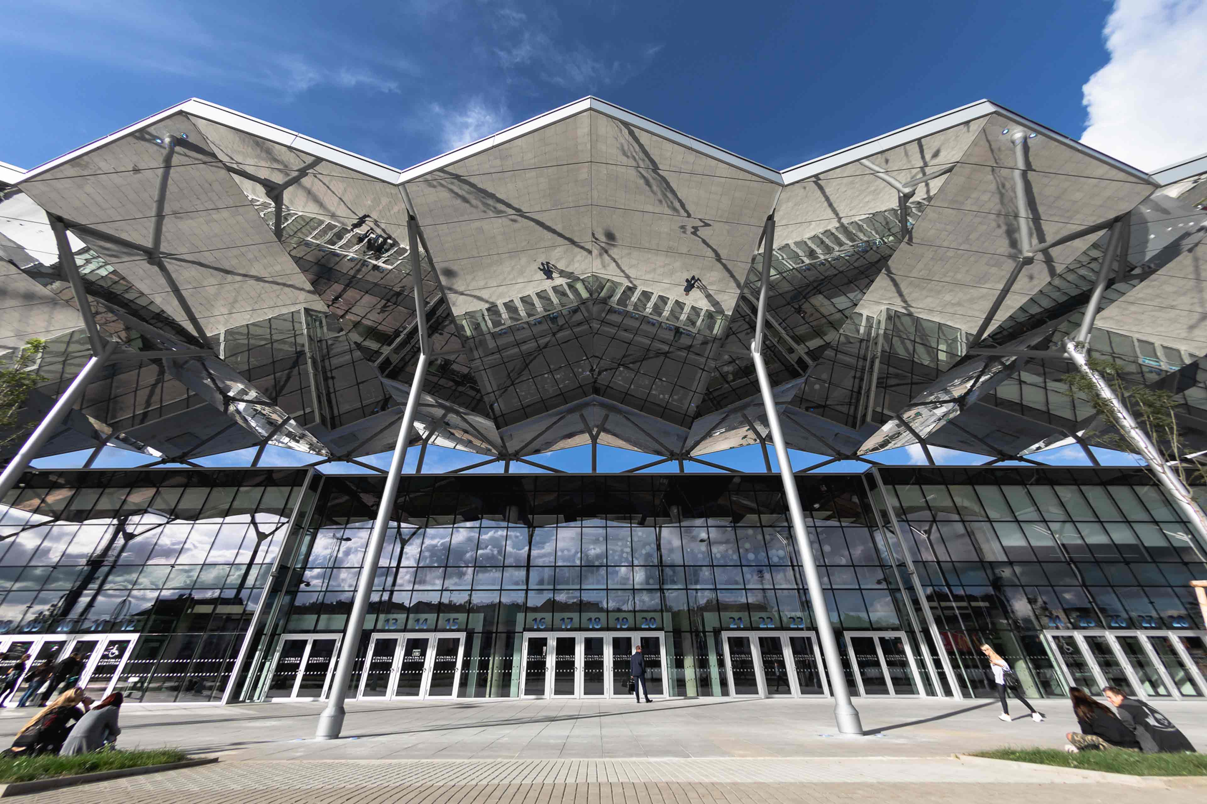
O2 universum
O2 universum
Discover Tomorrow‘s Dentistry
in the Heart of Magical Prague
in the Heart of Magical Prague
Room: O2
Date: 4.9.2026
Timings: 09:00 - 10:50
Abstract:
Artificial Intelligence is moving from research into everyday dental practice, supporting clinicians in making faster and more precise diagnostic decisions. By recognizing patterns in radiographs and clinical data, AI tools can assist in the detection of caries, periapical and endodontic pathology, periodontal bone loss, and other conditions that challenge routine diagnosis. This symposium will provide a clinically oriented overview of AI in diagnostics, with a focus on radiology and periodontology. Attendees will gain insight into how AI can reduce diagnostic variability, improve early detection, and enhance patient communication at the chairside.
Learning Objectives of The Session:
1. Describe the current capabilities and limitations of artificial intelligence in dental diagnostics
2. Evaluate the application of AI technologies in oral radiology and periodontology
3. Assess the practical integration of AI tools into clinical workflows
Moderator: Falk Schwendicke
Biography: (TBC)
Speaker 1: Dr Peter Fritz
Title: Artificial Intelligence in dentistry: foundations for ethical and efective practice
Abstract: While diagnostic AI enhances pattern detection, it falters precisely where dental training matters most: identifying dangerous outliers. This session explores how AI-powered tools can enhance diagnostic accuracy while preserving the clinical intuition and empathy that define expert care. We'll examine real-world uses in predictive, diagnostic, operational, professional and compassionate AI domains. We will focus on how AI complements, and when it complicates, clinical decision-making. Case studies highlight moments where AI strengthens confidence and where it challenges judgment. Attendees will gain frameworks for documenting AI influence in the patient record, supporting transparency, trust, and defensibility. We address how AI can reduce clinician bias while introducing new forms of algorithmic bias, and explore strategies for patient communication when AI findings diverge from human impression. Ultimately, diagnostic AI should not diminish the role of the dentist but should protect time for compassion and sharpen the human lens on patient care.
Learning Objectives
1. Identify diagnostic outliers where AI pattern recognition fails and clinical risk concentrates, applying the specialized training that dental education provides for recognizing atypical presentations and complex cases that fall outside standard algorithmic patterns and require expert clinical interpretation
2. Communicate AI diagnostic findings to patients effectively, explaining when technology supports clinical assessment while maintaining trust in professional expertise. Learn to discuss cases where AI and clinical judgment diverge, ensuring patients understand the role of both technology and human expertise in their care
3. Implement quality assurance protocols for AI diagnostics, including validation of AI findings against clinical judgment and documentation of diagnostic reasoning. Establish workflows that capture both AI contributions and clinical decision-making processes to ensure transparency, accountability, and defensible patient care records
4. Differentiate the five domains of AI in dentistry- Operational, Diagnostic, Predictive, Professional and Compassionate applications
Speaker 2: Antonin Tichy
Title: Demystifying Diagnostic AI in Dentistry: Making Informed Decisions
Abstract: Artificial intelligence (AI) is increasingly embedded in dental diagnostics, promising high accuracy across tasks such as the detection of caries or periapical radiolucencies. However, it can be difficult for clinicians to judge whether a tool is truly helpful or merely impressive on paper. This presentation will offer a practical, clinician-focused guide to understanding and evaluating diagnostic AI systems in dentistry. Through real-world examples, we will explore what “AI accuracy” actually means, why tools may perform well in some settings but not in others, and how clinicians can identify red flags in overly optimistic claims. Without delving into technical details, the presentation will provide clinicians with essential knowledge about diagnostic AI tools and equip them with key questions to ask when considering AI integration. The goal is to empower clinicians to make informed decisions about whether to trust and use diagnostic AI in daily practice.
Learning Objectives
1. Understand the role of diagnostic AI in common dental applications
2. Learn how to evaluate AI tools from a clinical perspective
3. Identify limitations and red flags in AI product claims
4. Build confidence in making informed decisions about diagnostic AI tools
Speaker 3: Assoc. Prof. Sergio Uribe
Title: To Make Artificial Intelligence Work in Dentistry, First Fix the Data
Abstract: Artificial intelligence in dentistry faces a critical barrier: data quality, not algorithmic sophistication. Current dental systems optimize for billing, not clinical outcomes, creating datasets that predict insurance approvals but not treatment success. Our data is broken. We document for billing, not outcomes. We lack standards. Systems don't communicate. In this presentation, we will discuss how to evaluate data quality, understand why standardization must precede AI deployment, and recognize their role in building clinical intelligence infrastructure.
Learning Objectives
1. Identify critical data requirements for training clinically useful AI models in their practice
2. Audit their clinical records for completeness, accuracy, and outcome documentation
3. Implement documentation standards that enable them to develop their own custom AI solutions
Room: D3-4
Date: 4.9.2026
Timings: 15:30 - 17:20
Abstract:
Artificial Intelligence is rapidly transforming healthcare, research, and society at large. With its promise come pressing challenges around ethical responsibility, data governance, and safeguarding sensitive information. This symposium brings together three leading experts to explore the complex intersections of AI ethics and data security. The session will move from ethical frameworks to technical safeguards, and finally to practical methods of implementation. Participants will gain insights into regulatory landscapes, cutting-edge approaches to secure AI systems, and concrete tools for integrating ethics and security into everyday AI projects. By combining conceptual, technical, and applied perspectives, the symposium equips attendees to critically assess risks while advancing responsible innovation.
Learning Objectives of The Session:
1. Evaluate the ethical, legal, and societal challenges of AI implementation
2. Identify and apply key principles of AI data security and governance
3. Develop practical strategies for operationalizing ethics and security in AI projects
Moderator: Falk Schwendicke
Biography: (TBC)
Speaker 1: Prof. Maxime Ducret
Title: The Diversity of Artificial Intelligence in Dentistry: Toward more Sustainability?
Abstract: Artificial intelligence (AI) is transforming dentistry through advances in diagnostics, treatment planning, education, and administration. Despite its clear potential, the adoption of AI remains fragmented and uneven. The landscape of AI applications in dentistry spans diverse users, tasks, and contexts, which offers challenges (but also opportunities) for sustainable integration. This presentation critically examines the conditions necessary for long-term, responsible deployment of AI in dentistry, identifying some key barriers such as siloed innovation, ethical uncertainty, and unequal infrastructure. Without cohesive engagement across stakeholder groups and collective actions, such as curriculum development, development of ethical frameworks, and regulatory initiatives, sustainable AI implementation might remain in distance. Finally, it outlines strategic steps for leveraging the diversity of the profession as a driver for sustainability.
Learning Objectives
1. Participants will be able to map current AI applications, across diagnostics, treatment planning, education, and administration, and recognize how diverse users and contexts shape adoption
2. Participants will learn to identify and assess the impact of siloed innovation and unequal infrastructure on long-term deployment
3. Participants will be able to identify the collective actions necessary to drive the sustainable implementation of AI in dentistry
Speaker 2: Prof. Dominik Gross
Title: AI Ethics with special consideration of data security
Abstract: When talking about AI ethics, data security and endangered personal rights quickly come to mind. This short presentation addresses these issues, but also focuses on other ethical problems that arise from the perspective of (dental) patients, such as the misrepresentation of certain patient groups in the data, the sometimes inadequately verified quality standards of new AI applications, unequal access to these applications, the threat of deskilling among doctors, a loss of trust in the dentist-patient relationship as a result of the prominent integration of AI, and the diffusion of responsibility (issue of ultimate responsibility). These and other aspects will be discussed.
Learning Objectives
1. Data Security
2. Unequal access to AI applications
3. Diffusion of responsibility
Speaker 3: Assoc. Prof. Kazunori Nozaki
Title: Standardizing Dental Patient Records: Current Discussions and Future Outlook
Abstract: As digital transformation advances across healthcare worldwide, dentistry faces challenges in ensuring that clinical information can be consistently recorded and shared across institutions and borders. Dental patient records remain heterogeneous, shaped by local regulations and clinical workflows, limiting their value for continuity of care and collaboration. This symposium focuses on ongoing discussions within ISO/TC 215 WG2 on internationally harmonized approaches to dental patient data, including emerging concepts such as an International Dental Patient Summary (IDPS). From a policy and standards perspective, the session explores why structured dental records are increasingly important and how alignment with broader health information standards can be considered while respecting national regulations and clinical diversity. Rather than presenting finalized standards or specific implementations, the symposium highlights the direction of current international standardization efforts and key considerations related to governance and interoperability, helping participants understand how these developments may influence everyday dental practice and international cooperation.
Learning Objectives
1. Understand why dental patient records are increasingly discussed as a policy and standards issue, and recognize the limitations of current heterogeneous dental records for continuity of care and cross-border collaboration
2. Recognize the goals and challenges of ongoing international discussions within ISO/TC 215 WG2, including emerging concepts such as harmonized dental patient summaries, and their relevance to diverse clinical and regulatory contexts
3. Appreciate how future harmonization of dental patient records may gradually influence everyday dental practice, professional responsibility, and international cooperation, without relying on specific systems or finalized implementations
Room: E1
Date: 5.9.2026
Timings: 15:30 - 17:20
Abstract:
Temporomandibular disorders (TMD) and orofacial pain demand evidence-based diagnosis and coordinated care. This symposium moves stepwise from diagnostic frameworks and conservative management through imaging-driven phenotyping to surgical decision-making and outcomes. Attendees will leave with practical tools to differentiate TMD from other orofacial pain conditions, apply diagnostic findings to patient-specific care plans, and understand when—and how—to escalate to minimally invasive or reconstructive procedures.
Learning Objectives of The Session:
1. Differentiate temporomandibular disorders from other orofacial pain conditions
2. Interpret imaging and clinical findings to support phenotype-guided management of TMD
3. Develop patient-centered treatment pathways for TMD and orofacial pain
Moderator: Vladimir Machoň
Biography: (TBC)
Speaker 1: Prof. Kai Yuan Fu
Title: Non-surgical disc reduction therapy for TMJ disc displacement without reduction
Abstract: Disc displacement without reduction (DDw/oR) is a common subtype of temporomandibular disorders (TMDs) that can lead to orofacial pain, limited mouth opening, and potential progression to degenerative joint disease (DJD). This lecture will address four key aspects: (1) the strong association between TMJ DJD and DDw/oR, (2) the non-surgical disc reduction protocol for DDw/oR, (3) factors influencing the success of non-surgical disc repositioning, and (4) the role of disc reduction in promoting condylar regenerative remodeling. Our clinical experience and research findings indicate that: (1) DDw/oR predisposes the condyle to degenerative changes in adolescents, a stage of active condylar remodeling, (2) the TMJ condyle exhibits significant self-repair capacity, enabling complete recovery from early-stage DJD through regenerative remodeling, (3) disc reduction treatment followed by anterior repositioning splint therapy is an effective and promising treatment for early TMJ DJD associated with disc displacement, (4) non-surgical disc reduction treatment for DDw/oR is most successful when initiated within 3 months of symptom onset.
Learning Objectives
1. To understand the specific detriments of TMD disc displacement without reduction in adolescents
2. To understand that early TMJ degenerative changes could be fully repaired after disc reduction treatment
3. To explain the procedures and treatment indications for Non-surgical disc reduction therapy
Speaker 2: Prof. David Psutka
Title: TMD and Orofacial Pain: From diagnostic Triage to Imaging Based Phenotyping and Surgical Pathways
Abstract: This will deal with evidence based indications for TMJ surgery including injections for masticatory myalgia and neuropathic pain, intra-articular injections (steroid, hyaluronic acid, platelet rich growth factors) arthrocentesis, arthroscopic surgery, open joint surgery and total joint replacement. Also reference to concomitant orthognathic surgery when indicated. Evidences based outcomes complication and revision surgery to be discussed.
Learning Objectives
1. To understand: indications for TMJ surgery
2. Types of TMJ surgery
3. Evidence based outcomes
4. Complications and revision surgery
Room: O2
Date: 5.9.2026
Timings: 09:00 - 10:50
Abstract:
Retaining natural teeth versus replacing them with implants requires calibrated judgment grounded in biology, biomechanics, and long-term risk. This symposium moves stepwise from defining prognostic markers for tooth preservation and the role of targeted antimicrobial therapy, through case-based criteria for selective extraction, to the scenarios in which implant therapy provides superior predictability and prosthetic stability. Attendees will leave with practical tools to balance periodontal recovery potential against implant advantages, translate diagnostic findings into individualized treatment plans, and determine when—and how—to shift from preservation to implant-supported rehabilitation.
Learning Objectives of The Session:
1. Assess prognostic factors for natural tooth retention versus extraction and replacement
2. Apply evidence-based criteria for selecting between conservative periodontal therapy, targeted antimicrobial strategies, selective extraction, and implant placement
3. Develop individualized treatment planning strategies that balance tooth preservation and implant rehabilitation
Moderator: Dr Jan Streblov
Biography: Dr. Streblov received his degree in dentistry in 2003 from the First Medical Faculty Charles University, Prague. During his studies he spent two semesters at the University of Rostock, Germany. 2004 - 2010 he worked as a dentist in the Prague Dental Implant Centre focusing on interdisciplinary dental treatment. He translated several textbooks for Quintessence Publishing serving as key translator for a variety of international speakers. In 2010 he co-founded the 3DK Dental Clinic in Prague with Dr. Martin Tomeček. He received certificates in Periodontology (2009, Czech Dental Chamber) and Implantology (2010, DGI - German Society for Implantology). Since 2010 he has presented numerous lectures in the fields of periodontology, implantology, esthetics, interdisciplinary treatment, autotransplantation of teeth and healing of oral tissues on national and international level. In the years 2019 - 2020 Dr. Streblov had the honour to serve as the president of Czech Academy of Dental Esthetics.
Speaker 1: Prof. Howard Glückman
Title: Complex Hard and Soft Tissue Defects in the Anterior Maxilla
Abstract: This lecture addresses the management of complex hard and soft tissue defects in the anterior maxilla, encompassing strategies from preservation to full restoration. We will delve into the intricacies of bone grafting and soft tissue grafting, discussing the optimal sources and techniques for harvesting these grafts. Our discussion will include the latest advancements in Partial Extraction Therapy (PET) and the socket shield technique, highlighting their roles in preventing alveolar ridge collapse post-extraction. By exploring these methods, participants will gain a comprehensive understanding of how to effectively manage and rehabilitate challenging anterior maxillary defects, ultimately enhancing patient outcomes and maintaining long-term oral health.
Learning Objectives
1. Understand the biological and clinical factors that contribute to complex hard and soft tissue defects in the anterior maxilla, and evaluate preservation-to-restoration strategies for optimal long-term outcomes
2. Analyse and apply evidence-based techniques in bone and soft tissue grafting, including indications, harvesting methods, and decision-making for selecting appropriate graft sources in challenging anterior cases
3. Evaluate the role of Partial Extraction Therapy (PET) and the socket shield technique in maintaining ridge integrity, and integrate these approaches into treatment planning to prevent post-extraction collapse and improve aesthetic and functional results
Speaker 2: Prof. Niklaus Lang
Title: To maintain the teeth- a treatment planning guide
Abstract: This paper presents a case of a patient with severe periodontitis, demonstrating how targeted and antimicrobial therapy can preserve the natural dentition and simultaneously facilitate prosthetic rehabilitation. Only teeth with attachment loss to the apex, external root resorption, or caries in an affected furcation were extracted. As a result, the reconstruction could be achieved with a minimal number of implants—significantly more conservatively than would have been the case with a radical extraction strategy. The successful management of the periodontal condition was largely due to the patient's high compliance and the consistent implementation of a structured antimicrobial treatment concept. The documented course of treatment illustrates that even severely periodontally compromised teeth may be preserved long-term with competent therapy, enabling both functional and aesthetic rehabilitation.
Learning Objectives
1. To develop a pre- therapeutic prognosis
2. To follow a systematic treatment sequence after comprehensive treatment planning. Maintaining natural abutments over implants
3. To establish an individually optimal treatment plan for esthetic and function under healthy conditions: infection control
Speaker 3: Dr Syngcuk Kim
Title: Contemporary Endodontic Therapy and Microsurgery: Preserving Natural Dentition in Complex Cases
Abstract: Contemporary endodontic therapy, combined with advanced endodontic microsurgery, has undergone significant evolution through the integration of three-dimensional (3D) file systems, operating microscopes, cone-beam computed tomography (CBCT), and bioceramic materials. These innovations have markedly improved treatment predictability and clinical outcomes. Current evidence and clinical experience indicate that success rates exceeding 90% can be achieved in retaining natural dentition—even in cases presenting with periapical lesions—provided that the tooth demonstrates minimal mobility (≤ Grade I) and absence of periodontal pocketing. This presentation will focus on the biological and clinical principles that support the preservation of natural teeth that might otherwise be deemed non-restorable or “condemned."
Learning Objectives
1. Identify the advanced instruments and biomaterials essential for modern endodontic and microsurgical success
2. Critically evaluate whether extraction and implant placement offer superior outcomes compared to preservation of natural dentition
3. Analyze the underlying reasons for the increasing prevalence of dental implants despite the high success rates of tooth retention through endodontic therapy
Room: O2
Date: 6.9.2026
Timings: 09:00 - 10:50
Abstract:
Vital pulp therapy has transitioned from niche to mainstream, offering a biologically grounded, minimally invasive alternative to root canal treatment—especially in deep caries management. This symposium moves stepwise: first, the histological reality of inflamed pulps and their healing potential; second, rigorously defined clinical protocols for partial/full pulpotomy in immature and mature permanent teeth; and third, the restorative-periodontal decisions that determine long-term success. Attendees will leave with an evidence-based algorithm that links diagnosis, haemostasis endpoints, material choices, and a robust coronal seal to predictable outcomes.
Learning Objectives of The Session:
1. Explain the biological and histological principles underlying vital pulp therapy
2. Apply evidence-based clinical protocols for vital pulp therapy
3. Evaluate restorative and long-term management factors that influence vital pulp therapy success
Moderator: Dr Daniel Černý
Biography: Daniel Cerny (*1974) has received his dental degree at the Charles University, Medical School in Hradec Kralove (1998). Doctorate degree earned at Palacky University in Olomouc in 2018 with the topic of adhesive aesthetic reconstruction. Previously an assistant professor at Charles University, Medical School in Hradec Kralove (1998-2007). Private practice in Hradec Kralove limited to endodontics and adhesive dentistry since 2001. Immediate Past President of the Czech Endodontic Association (CES) 2015-2023. Member of the Scientific Board of the Czech Dental Chamber. (since 2022) Co-founder and the first president of Czech Academy of Dental Esthetics (CADE) (2007-2009). Editorial board member of LKS journal (Czech Dental Chamber Journal) (2009-2013). International Member of AAE. Author/co-author of 36 articles in dental journals. Co-author of 4 book chapters. Lectures widely internationally on topics of endodontics and adhesive restorations with special focus on endo-restorations.
Speaker 1: Prof. Talal Al-Nahlawi
Title: The best root canal filing material ever; Vital Pulp Therapy
Abstract: The introduction of biomaterials in endodontics and the improvements happened, as well as proper understanding of inflamed dental pulp stage and related symptoms allowed for more vital pulp tissue preservation inside treated carious tooth minimizing the need for more tooth structure removal comparing with the traditional root canal treatment, the issue that will provide better circumstances for longer service duration of affected tooth inside oral cavity. The aim of this presentation is to highlight a series of 4 clinical cases of vital pulp therapy starting from a typical open apex management case till more complicated cases and management with long recalls.
Learning Objectives
1. Describe the conditions when we can preserve the dental pulp instead of performing pulpectomy
2. Describe how to perform partial pulpotomy
3. Describe how to perform full pulpotomy
4. Describe when to perform selective Vital Pulp Therapy
5. Describe follow up protocol and decide when the treatment is successful or not
Speaker 2: Prof. Alexis Gaudin
Title: From haemostasis to a hermetic seal
Abstract: Vital pulp therapy (VPT) aims to preserve pulpal vitality by controlling inflammation and providing conditions conducive to healing and long-term function. Beyond case selection and diagnosis, effective haemostasis and proper management of the pulp wound are key determinants of success, particularly when Hydraulic calcium silicate-based materials are used. This presentation focuses on the sequence from haemostasis to hermetic sealing in VPT, addressing the role of irrigants, haemostatic agents, and the timing of bioceramic placement. The effects of commonly used irrigants and haemostatic strategies on blood control, pulp tissue compatibility, and dentin surface characteristics are reviewed. Particular attention is given to the influence of residual bleeding on the setting reaction, adaptation, and sealing ability of bioceramics, as well as to the clinical relevance of immediate versus delayed placement. Practical, evidence-based guidance is provided to enhance the predictability of VPT outcomes.
Learning Objectives
1. Understanding the biological importance of haemostasis in vital pulp therapy: Rapid and controlled haemostasis is not merely a technical step but a biological indicator of pulp health and healing potential. Prolonged or uncontrolled bleeding may suggest irreversible inflammation and is associated with poorer outcomes
2. Identifying appropriate irrigants and haemostatic agents for VPT: A range of irrigants and haemostatic agents are used during VPT, each with distinct biological and chemical properties. A clear understanding of these agents enables clinicians to select protocols that balance bleeding control, biological compatibility, and material performance
3. Determine the optimal timing for bioceramic placement following haemostasis: The timing of bioceramic placement is critical to achieving a stable hermetic seal. Placement onto a bleeding surface or in the presence of residual blood proteins can alter the hydration, setting reaction, and mechanical properties of calcium silicate-based materials. Conversely, excessive delay or overly aggressive drying may compromise pulp vitality
Speaker 3: Dr Domenico Ricucci
Title: Biologically Driven Pulpotomy: From Histologic Infection Patterns to Clinical Decision-Making
Abstract: After outlining the histologic and histobacteriologic events that lead to the formation of an initial focal area of pulpal necrosis following bacterial penetration, the subsequent progression of tissue degeneration is analyzed. Based on the position of the advancing infectious front, a biologically driven treatment protocol is proposed. The clinical management consists of the stepwise excision of diseased and infected pulp tissue, followed by meticulous evaluation of the exposed wound surface after achievement of hemostasis. Completion of the pulpotomy is indicated when the wound appears homogeneous and uniformly blood-filled, without yellowish or brownish discolorations suggestive of liquefaction or coagulative necrosis. The exposed pulp tissue is then sealed with a bioceramic material to promote healing and hard tissue formation. The clinical and histologic outcomes of this approach are discussed in detail, together with an analysis of the biological and technical factors associated with treatment failure.
Learning Objectives
1. Understand the histologic and histobacteriologic progression of pulpal infection, from initial bacterial penetration to the formation and expansion of focal necrosis
2. Identify clinical criteria for biologically guided pulpotomy, including assessment of the infection front, stepwise pulp tissue removal, and interpretation of wound appearance after hemostasis
3. Evaluate the biological rationale, clinical outcomes, and causes of failure in vital pulp therapy, with particular emphasis on the role of bioceramic materials in promoting healing and regeneration
Room: E1
Date: 7.9.2026
Timings: 15:30 - 17:20
Abstract:
3D imaging has transformed orthodontics and orthognathic surgery. This symposium spans CBCT indications and incidental findings, imaging-guided orthodontic mechanics, and 3D planning for orthognathic surgery. Participants will learn when CBCT is justified, how to systematically interpret volumes, and how radiology improves biomechanics and surgical outcomes.
Learning Objectives of The Session:
1. Apply evidence-based principles for the appropriate use and interpretation of CBCT in orthodontics and orthognathic surgery
2. Evaluate the role of 3D imaging in guiding orthodontic biomechanics and treatment planning
3. Integrate virtual surgical planning and interdisciplinary collaboration in orthognathic treatment
Moderator: Assoc. Prof. Marek Ivo
Biography: Doc. MUDr. Ivo Marek, PhD, graduated with a degree in Dentistry from the Faculty of Medicine at Palacky University in Olomouc in 1990. He completed a three-year training program for Orthodontics in 1999 and finished his postgraduate study of Orthodontics at the Orthodontic Department of Dental School at Palacky University in Olomouc in 2007, where he earned his PhD. Dr. Marek runs his Private Dental Clinic in Breclav, where he focuses on interdisciplinary dental care, tooth autotranplantation and collaboration of orthodontists with periodontists and implantologists. He also was the first orthodontist in the Czech Republic to start using Invisalign aligners in 2007. Dr. Marek works as an Assistent Professor at the Orthodontic Department of Dental School, Palacky University, in Olomouc, in both graduate and postgraduate study programs. In addition, he is a part-time teacher at Charles University's Faculty of Medicine, in Prague. In 2025, he habilitated and earned the title of associa
Speaker 1: Prof. Ingrid Różyło-Kalinowska
Title: Beyond the Smile: Incidental Findings That Could Change Your Treatment Plan
Abstract: Diagnostic imaging has become an indispensable tool in orthodontics, providing detailed insights into the complex anatomical structures of the craniofacial region. Out of many different imaging modalities it is the Cone-Beam Computed Tomography (CBCT) which is more and more applied. It not only presents morphology and allows diagnosing of abnormalities, but is also integrated with intraoral scanning provides simulation of treatment outcomes. Beyond enabling precise assessment of dental and skeletal discrepancies, radiological examinations often reveal incidental findings - unexpected pathologies or anomalies—that can significantly influence treatment planning and patient management. The aim of the lecture is to present incidental findings not related to dental abnormalities that can lead to changes in treatment plans. The following findings will be discussed: airway, cervical vertebrae, soft tissue calcifications and sella turcica. Recognizing the importance of thorough interpretation of diagnostic imaging studies in orthodontics is essential for comprehensive patient care and for avoiding potential overlooked health issues.
Learning Objectives
1. To learn imaging findings related to soft tissue calcifications in CBCT in dental practice
2. To understand the importance of incidental findings which may lead to changes in dental treatment plan
3. To learn which incidental findings may reveal diseases that will require medical attention and postponing of dental treatment
Speaker 2: Dr Suliman Shahin
Title: Virtual Surgical Planning: Integrating Clear Aligner Therapy and Surgery in the CBCT Era
Abstract: Virtual surgical planning has transformed the interdisciplinary management of orthodontic and orthognathic cases, particularly when combined with clear aligner therapy. This presentation focuses on the integration of CBCT-based imaging into digital orthodontic-surgical workflows to enhance diagnostic accuracy, treatment coordination, and outcome predictability. Key topics include CBCT-driven facial and skeletal analysis, digital setup validation, aligner-based presurgical decompensation, and surgical simulation. The role of CBCT in facilitating communication between orthodontists, surgeons, and radiologists will be highlighted, emphasizing precision in planning and execution. Clinical cases will demonstrate how virtual planning improves soft- and hard-tissue outcome prediction, reduces treatment inefficiencies, and supports patient-centered decision-making. Limitations, sources of error, and practical challenges in real-world implementation will also be discussed, providing attendees with a realistic and clinically applicable framework for interdisciplinary virtual planning in the CBCT era.
Learning Objectives
1. Understand the role of CBCT in virtual surgical planning for combined clear aligner and orthognathic treatments
2. Learn how to integrate orthodontic digital setups with surgical simulation to improve interdisciplinary coordination
3. Identify clinical benefits, limitations, and potential sources of error in CBCT-based virtual surgical workflows
Speaker 3: Dr Carlos Villegas
Title: Proportional condylectomy: An alternative to orthognathic surgery without orthognathic surgery
Abstract: The management of facial asymmetries has been a challenge for pediatric dentists in childhood and for orthodontists in adolescence. Therefore when early correction is not successful orthognathic surgery is required in most cases. Proportional condylectomy is a treatment alternative that, when combined with orthodontic treatment using skeletal anchorage (TADS), has proven effective for the successful correction of asymmetry. The scientific evidence and patient management with this treatment alternative will be presented.
Learning Objectives
1. Learn to identify cases that could benefit from this treatment option
2. Understand the conservative management of facial asymmetry step by step using skeletal anchorage
3. Consider this treatment alternative at an early age to avoid overtreatment of patients


© 2026 | Operator: GUARANT International spol. s r.o. | E-mail: guarant@guarant.cz | Powered by APPTIDE
Českomoravská 19, 190 00 Prague 9, Czech Republic
Reg. No.: 45245401 | VAT No.: CZ45245401 | Recorded in the Commercial Register, file No. 7144.